
Code |
БИМКЧА |
632607 |
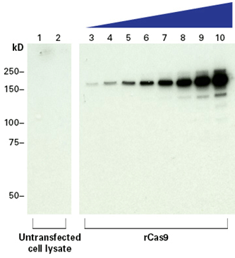
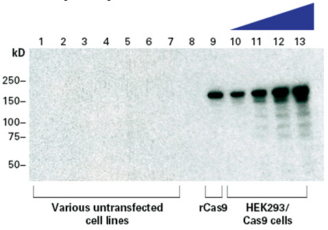
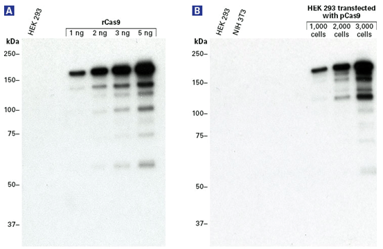
100 Ѕьl Guide-it Cas9 Polyclonal Antibody (1 Ѕьg/Ѕьl) |
632606 |
3 x 100 Ѕьl Guide-it Cas9 Polyclonal Antibody (1 Ѕьg/Ѕьl) |
632628 |
100 Ѕьg Guide-it Cas9 Monoclonal Antibody (Clone TG8C1) (1 Ѕьg/Ѕьl) |
632627 |
3 x 100 Ѕьg Guide-it Cas9 Monoclonal Antibody (Clone TG8C1) (1 Ѕьg/Ѕьl) |